Medical Diagnostic Imaging
- Introduction:
- Diagnostic imaging, also called medical imaging, the use of electromagnetic radiation and certain other technologies to produce images of internal structures of the body for the purpose of accurate diagnosis.
- Diagnostic imaging refers to a variety of non-invasive methods for identifying and monitoring diseases or injuries via the generation of images representing internal anatomic structures and organs of the patient’s body. The detailed images produced by these procedures are used to further inform the patient and physician about the anatomic organization and functional working of the inner organs and structure of the patient’s body. Diagnostic imaging is an informational tool that expands the knowledge of physicians, people and patients and the practice of medicine.
- Radiologists and other physicians interpret the resulting images to diagnose various medical illness or injury so that patient treatment and therapy can be specifically planned and implemented. Diagnostic imaging is also used to guide surgical planning and is often used to follow surgery and/or monitor the outcomes of therapeutic procedures.
- Diagnostic imaging denotes that new innovative techniques like ultrasound (US), magnetic resonance (MR) and computed tomography (CT) are now also performed in radiology centers and departments along with traditional x-ray or radiology
- Diagnostic Imaging is a Key Tool in the Early Diagnosis and Prevention of Disease

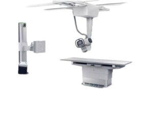
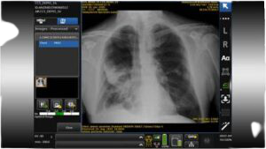
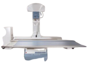
Fixed Radiography Systems
- imaging into your radiography room, helping you resolve inconclusive x-rays with minimal added radiation. Our cost-effective technology helps improve patient experience by providing additional information that could help make care decisions faster.
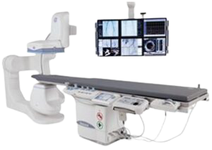
Fluoroscopy Systems
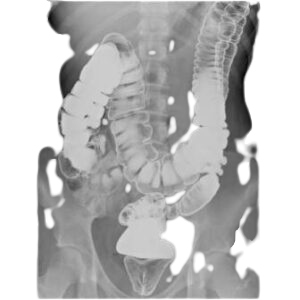
- is a type of medical imaging that shows a continuous X-ray image on a monitor, much like an X-ray movie. During a fluoroscopy procedure, an X-ray beam is passed through the body. An imaging technique that uses X-rays to obtain real-time moving images of the interior of an object. In its primary application of medical imaging, allows a physician to see the internal structure and function of a patient, so that the pumping action of the heart or the motion of swallowing, for example, can be watched. This is useful for both diagnosis and therapy and occurs in general radiology, interventional radiology, and image-guided surgery. In its simplest form, a fluoroscope consists of an X-ray source and a fluorescent screen, between which a patient is placed.

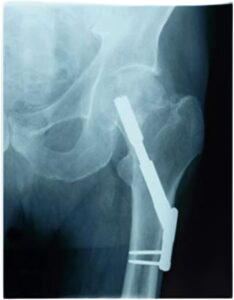
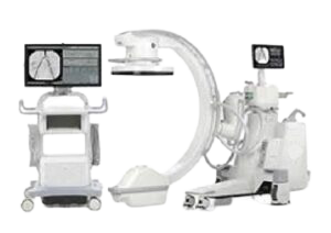
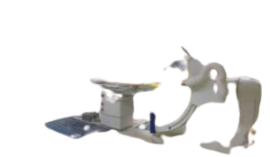
Surgical Imaging
- A C–arm is an imaging scanner intensifier. The name derives from the C-shaped arm used to connect the x-ray source and x-ray detector to one another. C–arms have radiographic capabilities, though they are used primarily for fluoroscopic intraoperative imaging during surgical, orthopedic and emergency care procedures Achieve optimum visual detail during vascular procedures in a hospital. See more anatomy with exceptional detail at low dose With advanced technology, intelligent workflow, and dose control, is the future of mobile surgical imaging. This system is ideally suited for a range of surgical needs including vascular, cardiac, orthopedics, gastrointestinal, endoscopic, urologic, critical care, pain management and emergency procedures.
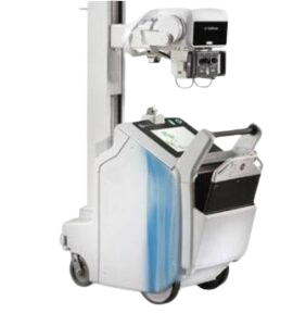
Mobile Radiography Systems
- Mobile radiography using transportable radiographic equipment allows imaging services to be brought to the patient. In contrast to the large stationary machines found in radiographic rooms, compact mobile radiography units can produce diagnostic images in virtually any location. Mobile radiography is commonly performed in patient rooms, emergency departments, intensive care units, surgery and recovery rooms, and nursery and neonatal units. Some machines are designed for transport by automobile or van to nursing homes, extended care facilities, or other off-site locations requiring radiographic imaging services.
Advanced Digital Radiography Systems
- From standard radiography as the first diagnostic test for most chest, orthopedic and trauma cases. Advanced medical imaging is often ordered for complex cases that radiography cannot solve. These additional imaging modalities may result in higher radiation exposure, higher imaging costs, and a delay in diagnosis & treatment that could also mean more anxiety and discomfort for the patient. Advanced Digital Radiography imaging into your radiography room, helping you resolve inconclusive x-rays with minimal added radiation. Cost-effective technology helps improve patient experience by providing additional information that could help make care decisions faster. Obtain multiple cross-section images of the anatomy in a single sweep at a low dose, including chest, abdomen, extremities, spine, skull & sinuses.
Resolve inconclusive x-rays in orthopedic & trauma cases efficiently . Achieve superior lung nodule detection sensitivity compared to conventional chest x-ray
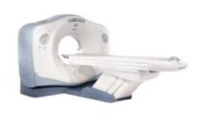
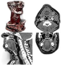
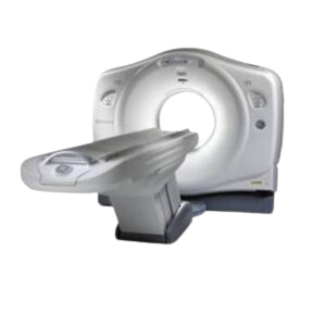
Computed Tomography
- Expedite patient care and enhance laboratory efficiency with our automation systems. Deliver rapid, consistent results to facilitate diagnosis and treatment—potentially improving patient care, emergency room times and patient/clinician satisfaction. Using Pre- and Post-analytical Automation Solutions helps ensure consistent turnaround times and eliminates opportunities for error, improving accuracy of results and supporting more informed decision-making when it comes to patient care combining disciplines on a single rack helps meet busy laboratory demands and stockyards—specialized refrigerated outlet units—allow sample storage for a configurable period of time based on laboratory workflow
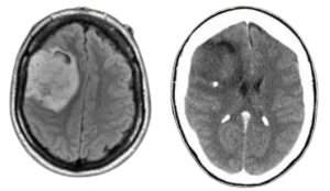
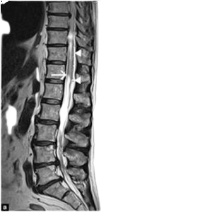
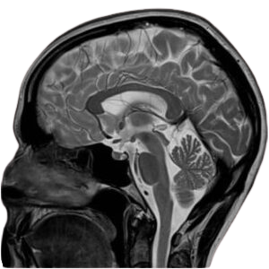
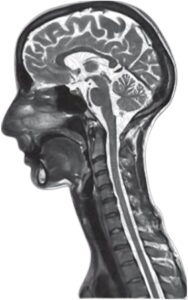
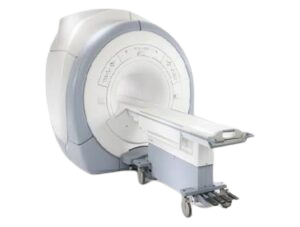
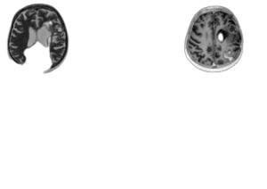
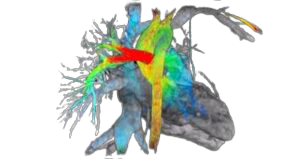
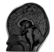
Magnetic Resonance Imaging
- Magnetic resonance imaging is a medical imaging technique used in radiology to form pictures of the anatomy and the physiological processes of the body. MRI scanners use strong magnetic fields, magnetic field gradients, and radio waves to generate images of the organs in the body. MRI does not involve X-rays or the use of ionizing radiation, which distinguishes it from CT and PET scans. MRI is widely used in hospitals and clinics for medical diagnosis, staging of disease and follow-up without exposing the body to radiation. An MRI may yield different information compared with CT , MRI is excellent in providing soft tissue details .
- By Field Strength
- Very-high—field MRI systems (4T and above)
- High-field MRI systems (1.5T to 3T)
- Low-to-mid-field MRI systems (less than 1.5T)
By Application
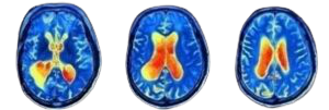
- Neuro MRI focuses on imaging and therapies to diagnose and to treat disorders of the adult and pediatric brain, spine, neck, and central and peripheral nervous system
- Cardiac Magnetic Resonance Imaging (MRI) is used to detect or monitor cardiac disease and to evaluate the heart’s anatomy and function in patients with both heart disease present at birth and heart diseases that develop after birth.
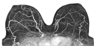
- Breast MRI is the most sensitive method for detection of breast cancer. Depending on international health regulations, it is either applied for screening of women
- vascular MRI scanning is a noninvasive imaging technique that does not involve exposure to ionizing radiation. … The contrast material used in a cardiac MRI scan is less likely to produce an allergic reaction than the iodine-based contrast materials used for conventional x-rays and CT scanning.
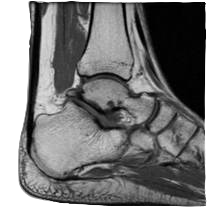
- Extremity MRI is a type of scan used specifically for diagnostic imaging of the arm, leg, hand, or foot.
- pediatric MRI is the imaging modality of choice in a vast array of pediatric conditions, particularly in neurologic, musculoskeletal, and some cardiovascular diseases
By Architecture
- Closed MRI systems
- Standard bore MRI
- Wide bore MRI
- Open MRI systems
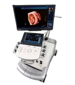
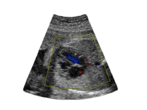
Ultrasound
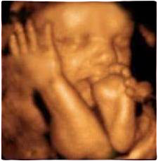
- Ultrasound imaging uses sound waves to produce pictures of the inside of the body. It is used to help diagnose the causes of pain, swelling and infection in the body’s internal organs and to examine a baby in pregnant women and the brain and hips in infants. It’s also used to help guide biopsies, diagnose heart conditions, and assess damage after a heart attack. Ultrasound is safe, noninvasive, and does not use ionizing radiation.
- By Application:
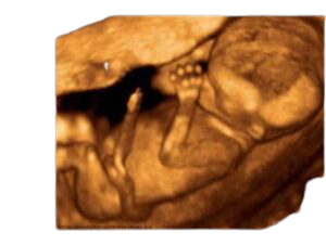
- Obstetrics / Gynecology ultrasound uses sound waves to produce pictures of a baby (embryo or fetus) within a pregnant woman, as well as the mother’s uterus and ovaries , and is the preferred method for monitoring pregnant women and their unborn babies. A Doppler ultrasound study – a technique that evaluates blood flow in the umbilical cord, fetus or placenta – may be part of this exam.
Ultrasound By Application Cont’d
- General Radiology Ultrasound of heart and blood vessels, including the abdominal aorta and its major branches Liver Gallbladder Spleen Pancreas Kidneys Bladder Uterus, ovaries, and unborn child (fetus) in pregnant patients Eyes Thyroid and parathyroid glands Scrotum (testicles)
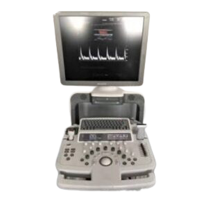
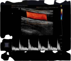
- Cardiac Ultrasound, also known as echocardiography, concerns the ultrasound imaging of a very fast moving complex organ positioned deep within the body – the heart. The analysis of images is also heavily technology driven as resolving the diseases of the heart require a rather detailed representation of everything that moves (cardiac muscles, valves, blood) within the heart.

Cardiac Cath Lab
- Cardiac catheterization is a procedure used to diagnose and treat certain cardiovascular conditions. During cardiac catheterization, a long thin tube called a catheter is inserted in an artery or vein to the heart. Using this catheter, doctors can then do diagnostic tests as part of a cardiac catheterization. Some heart disease treatments, such as coronary angioplasty and coronary stenting, also are done using cardiac catheterization

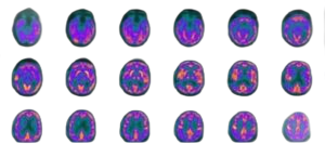
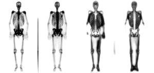
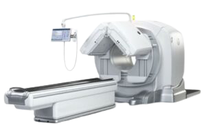
Molecular Imaging and PET Systems
- A positron emission tomography (PET) scan is an imaging test that helps reveal how your tissues and organs are functioning. A PET scan uses a radioactive drug (tracer) or radiopharmaceuticals to show this activity. This scan can sometimes detect disease before it shows up on other imaging tests. A PET scan is useful in revealing or evaluating several conditions, including many cancers, heart disease and brain disorders. Often, PET images are combined with CT or MRI scans to produce special views. A PET scan measures important body functions, such as metabolism. It helps doctors evaluate how well organs and tissues are functioning. Molecular imaging technologies include :
- Nuclear Medicine
- PET/CT
- PET Radiopharmacy

Cyclotron
- a type of particle accelerator, accelerates charged particles outwards from the center along a spiral path using a high frequency voltage and a magnetic field. It is used in nuclear physics research and for the production of radio isotopes in imaging and radiation therapy. Tracer production and PET/CT go hand in hand. Because of the relatively short half-lives of PET radioisotopes, it’s a race against the clock to produce a radioactive isotope, synthesize it into a radiopharmaceutical and get it to the patient. To go in search of true discovery with PET, access to a cyclotron is necessary,

Nuclear Medicine
- Nuclear medicine imaging uses small amounts of radioactive materials called radiotracers that are typically injected into the bloodstream, inhaled or swallowed. The radiotracer travels through the area being examined and gives off energy in the form of gamma rays which are detected by a special camera and a computer to produce images of the inside of your body. Nuclear medicine imaging provides unique information that often cannot be obtained using other imaging procedures and offers the potential to identify disease in its earliest stages. Nuclear medicine imaging non-invasively provides functional information at the molecular and cellular level that contributes to the determination of health status by measuring the uptake and turnover of target-specific radiotracers in tissue. These functional processes include tissue blood flow and metabolism, protein—protein interactions, expression of cell receptors in normal and abnormal cells, cell—cell interactions, neurotransmitter activity, cell trafficking and homing, tissue invasion, and programmed cell death. By providing information on these processes, nuclear medicine imaging offers a broad array of tools for probing normal and disease-related states of tissue function and response to treatment.
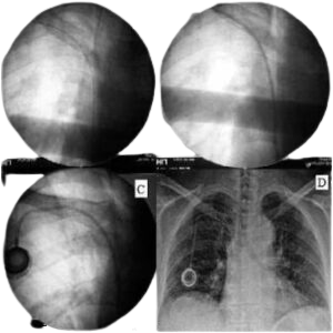
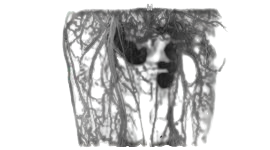
Angiography Systems
- angiogram is a procedure that uses X-ray imaging to see your heart’s blood vessels. The test is generally done to see if there’s a restriction in blood flow going to the heart. Coronary angiograms are part of a general group of procedures known as heart (cardiac) catheterizations. A coronary angiogram, which can help diagnose heart conditions, is the most common type of cardiac catheterization procedure. During a coronary angiogram, a type of dye that’s visible by an X-ray machine is injected into the blood vessels of your heart. The X-ray machine rapidly takes a series of images (angiograms), offering a look at your blood vessels. If necessary, your doctor can open clogged heart arteries (angioplasty) during your coronary angiogram.
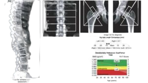
Bone Mineral Densitometry
- A bone mineral density test uses X-rays to measure the amount of minerals — namely calcium — in your bones. This test is important for people who are at risk for osteoporosis, especially women and older adults. The test is also referred to as dual energy X-ray absorptiometry (DXA) Dual energy X-ray Absorptiometry (DXA) is considered the most highly developed and most thoroughly validated technique for assessing bone mineral density.
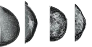
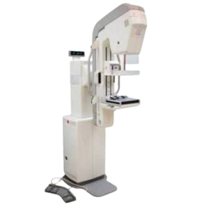
Mammography
- A mammogram is an x-ray picture of the breast. It can be used to check for breast cancer in women who have no signs or symptoms of the disease. It can also be used if you have a lump or other sign of breast cancer. Screening mammography is the type of mammogram that checks you when you have no symptoms , mammogram images of outstanding detail and superior diagnostic accuracy and with the same low dose as a 2D exam designed to deliver superior diagnostic accuracy for the detection of breast cancer during a mammogram
The Mobetron is the first mobile, self-shielded electron-beam linear accelerator (LINAC) machine designed to deliver
Intraoperative Radiation Therapy (IORT) to cancer patients during surgery. This innovation brings safe, reliable and portable
radiation to the operating room without the need for costly shielding renovations or retrofits. And in turn, Mobetron underscores
IntraOp’s commitment to providing a solution designed to maximize cost effectiveness and reduce risk for cancer
centers and their patients alike.
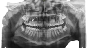
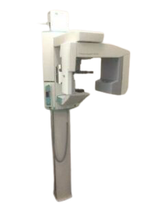
Panoramic Radiography
- also called panoramic x–ray, is a two-dimensional (2-D) dental x–ray examination that captures the entire mouth in a single image, including the teeth, upper and lower jaws, surrounding structures and tissues. Panoramic dental x-ray uses a very small dose of ionizing radiation to capture the entire mouth in one image. It is commonly performed by dentists and oral surgeons in everyday practice and may be used to plan treatment for dentures, braces, extractions and implants .
.
.
.
.
.
.